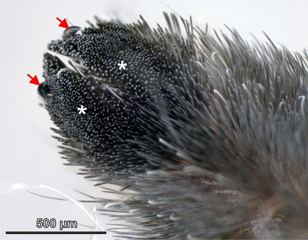
Scanning Electron Microscopy (SEM) image of the bases of pretarsal (ie, on least expensive part of leg) adhesive hairs. (A) On the left are the hair shafts of the adhesive hairs closest to the exoskeleton. SEM images of the microstructure of the adhesive hairs ( setae). (A) Side view showing the up to 1.8 mm long hair shaft (not revealed in complete length) and the suggestion area covered with microtrichia (minute hair-like structures on the hairs appropriate).( A) Downward-facing surface area of the tuft of hairs around the claws on the pretarsus (white asterisks mark the 2 lobes of the hair tuft), which consists of thousands of densely jam-packed hairs.
Scanning Electron Microscopy (SEM) image of the bases of pretarsal (ie, on lowest part of leg) adhesive hairs. (A) On the left are the hair shafts of the adhesive hairs closest to the exoskeleton. At their insertion, the hair shaft becomes thinner and a stopper-like structure on the exoskeleton connects and satisfies to it. (b) Further magnification of the very same area: the asterisk marks the pivot point where hairs can flex upwards. Proximal vs. distal here suggests away from vs. towards the claw on the suggestion of the leg. Credit: B Poerschke, SN Gorb and F Schaber
Engineers impressed by the functional excellent variety of hairs on spider legs.
Simply how do spiders walk directly– and even upside-down across– numerous different kinds of surface areas? Answering this concern could open new chances for creating powerful, yet reversible, bioinspired adhesives. Scientists have been working to much better understand spider feet for the previous a number of decades. Now, a new research study in Frontiers in Mechanical Engineering is the first to show that the attributes of the hair-like structures that form the adhesive feet of one species– the roaming spider Cupiennius salei– are more variable than previously believed.
SEM pictures of the microstructure of the adhesive hairs ( setae). (A) Side view showing the up to 1.8 mm long hair shaft (not revealed in complete length) and the pointer region covered with microtrichia (minute hair-like structures on the hairs proper). (B) Top view of the scopula pad (a dense tuft of hairs) on the lower side of the pretarsus. Covering the idea area of the hairs are spatula-shaped microtrichia, which stick to the substrate throughout walking. (C) Higher magnification picture of the spatula-shaped microtrichia. Credit: B Poerschke, SN Gorb and F Schaber
” When we began the experiments, we anticipated to discover a specific angle of best adhesion and similar adhesive properties for all of the individual attachment hairs,” states the group leader of the research study, Dr Clemens Schaber of the University of Kiel in Germany. “But surprisingly, the adhesion forces mainly varied in between the private hairs, e.g. one hair adhered best at a low angle with the substrate while the other one carried out best near perpendicular.”
The feet of this species of spider are comprised of near 2,400 small hairs or setae (one hundredth of one millimeter thick). Schaber, and his colleagues Bastian Poerschke and Stanislav Gorb, gathered a sample of these hairs and then determined how well they adhered to a range of smooth and rough surface areas, consisting of glass. They also took a look at how well the hairs carried out at numerous contact angles.
Various kinds of hair collaborate
Unexpectedly, each hair revealed special adhesive properties. When the group took a look at the hairs under a powerful microscope, they likewise discovered that every one proved different– and previously unrecognized– structural plans. The group believes that this variety might be key to how spiders can climb up many surface area types.
This present work studied just a small number of the thousands of hairs on each foot, and its beyond the scope of existing resources to think about studying them all. The group anticipates that not all of the hairs are special, and that it may be possible to discover clusters or duplicating patterns rather.
Bioinspired applications possible
” Although it is still extremely tough to fabricate nanostructures like those of the spider– and specifically to attain the stability and dependability of the natural products– our findings can even more optimize existing designs for residue-free and reversible synthetic adhesives,” says Schaber. “The principle of different shapes and positionings of adhesive contacts as found in the spider attachment system can enhance the attachment capability of bioinspired products to a broad variety of substrates with different homes.”
( A) Downward-facing surface area of the tuft of hairs around the claws on the pretarsus (white asterisks mark the 2 lobes of the hair tuft), which consists of thousands of largely packed hairs. (B) Side view of the hair tuft on the pretarsus sticking to a glass slide. Note how the ideas of the hairs have ends up being bent.
Recommendation: “Adhesion of Individual Attachment Setae of the Spider Cupiennius salei to Substrates With Different Roughness and Surface Energy” by Bastian Poerschke, Stanislav N. Gorb and Clemens F. Schaber, 11 June 2021, Frontiers in Mechanical Engineering.DOI: 10.3389/ fmech.2021.702297.


